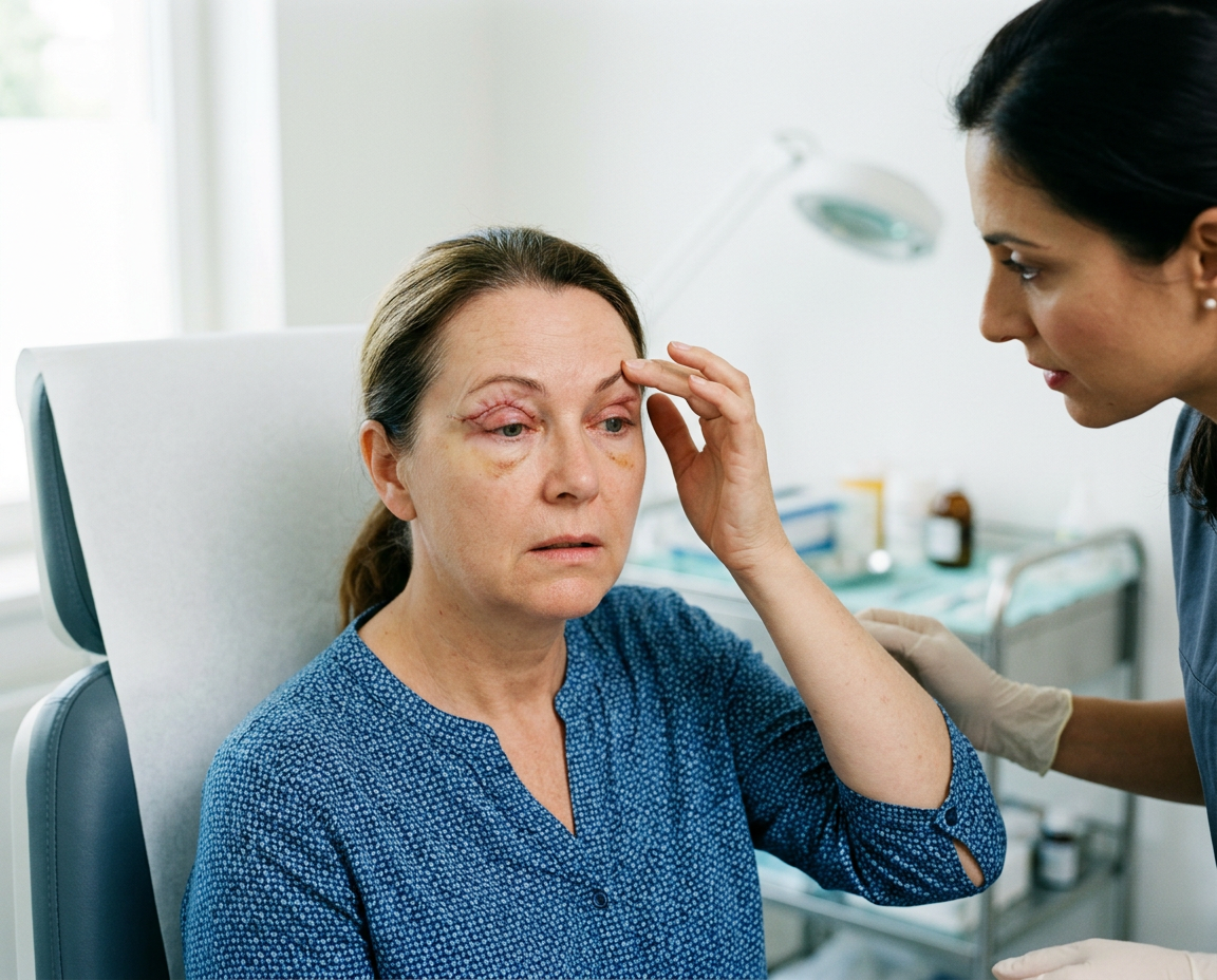
סיבוכים בהרמת עפעפיים: מה חשוב לדעת לפני הניתוח
סיבוכים בהרמת עפעפיים נחשבים לרוב לנדירים כאשר הניתוח מבוצע על

|
Getting your Trinity Audio player ready...
|
Barley in the eye is common in the eyelids and it affects many people in different age groups. It is characterized by a small and painless lump or swelling that forms on the upper or lower eyelid due to inflammation of the meibomian glands. These glands are responsible for producing the oily layer of tears that lubricates the eye. When these glands become blocked, the trapped oil can lead to the formation of corns in the eye. Although it is usually benign, barley in the eye can cause discomfort and cosmetic disturbance, prompting people to seek medical attention. Understanding stye treatment options and management strategies for stye is essential for both patients and healthcare providers to ensure effective resolution and prevention of recurrence of the condition.

Eyelids play an essential role in protecting and maintaining the health of the eyes. They consist of several layers, including the skin, muscle and connective tissue, which house the meibomian glands. These glands are located inside the tarsal plate of the eyelids and are responsible for the secretion of the fatty component of the tears. The tears themselves consist of three layers: the outer lipid layer, the middle aqueous layer and the inner mucin layer. The lipid layer, produced by the meibomian glands, prevents the evaporation of the aqueous layer and maintains the stability of the tear layer.
In the pathogenesis of barley in the eye, obstruction of the meibomian gland duct leads to accumulation of glandular secretions. This blockage can be due to various factors, including chronic inflammation, gland dysfunction or bacterial infection. As the retained secretions accumulate, they create localized swelling within the eyelid, leading to a granulomatous inflammatory reaction. This reaction is characterized by the presence of macrophages, lymphocytes and other cells, which try to repel the foreign substance and resolve the blockage.
Distinguishing barley in the eye from other eyelid lesions, such as hordeolum, is essential for proper management. While both conditions involve the meibomian glands, hordeolum is an acute bacterial infection characterized by pain, redness, and tenderness. In contrast, barley in the eye is usually not painful and results from chronic inflammation rather than an acute infection. Accurate diagnosis and understanding of the underlying anatomy and pathophysiology are essential to determining the appropriate treatment approach for barley in the eye.
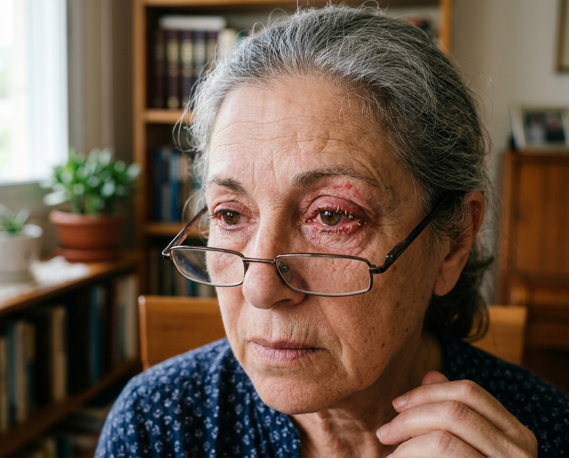
Barley in the eye usually appears as a local painless swelling of the upper or lower eyelid. Patients often notice the gradual development of a firm, round lump that can vary in size from a small pea to a larger, more palpable lump. Unlike hordeolum, which is often tender and associated with redness, barley in the eye usually lacks significant pain. However, barley in a larger eye can cause discomfort or a feeling of heaviness in the eyelid.
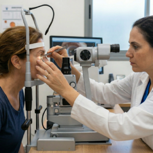
On physical examination, barley in the eye appears as a lump, not soft inside the tarsal plate. It may be visible from the outer surface of the eyelid or palpable by inverting the eyelid. If the barley in the eye is large enough, it can put pressure on the cornea, which can lead to blurred vision or astigmatism. Occasionally, a secondary infection can occur, turning the barley in the eye into an acute and painful inflammatory lesion.
Patients may also report a history of recurrent cataracts, especially if they have underlying disorders such as blepharitis, rosacea, or meibomian gland dysfunction. It is important to take a comprehensive history and perform a thorough examination to rule out other potential causes of eyelid swelling and confirm the diagnosis of barley in the eye.
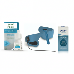
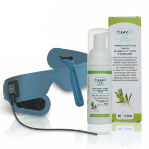
The initial management of barley in the eye often involves conservative treatment measures aimed at reducing inflammation and promoting drainage of the obstructed meibomian gland. One of the most recommended methods is applying warm compresses to the affected eyelid. This helps to liquefy the trapped secretions and facilitates drainage through the opening of the gland. Apply warm compresses for 10-15 minutes, three to four times a day, to achieve the desired effect.
In addition to warm compresses, maintaining good eyelid hygiene is essential. This includes gentle cleansing of the eyelid margin with a mild, non-irritating cleanser or eyelid scrub. Proper eyelid hygiene can help prevent the recurrence of stye in the eye by reducing the risk of gland blockage and inflammation.
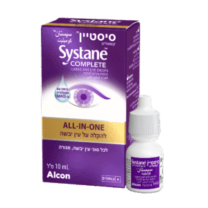
Topical medications, such as antibiotic or steroid eye drops, may be prescribed to manage associated inflammation or secondary infection. These medications can help reduce swelling and promote resolution of the barley in the eye. However, their effectiveness may be limited in cases where the barley in the eye has already formed a stable structure.
In some cases, oral antibiotics may be considered, especially if there is evidence of a secondary bacterial infection or if the patient has an underlying medical condition such as rosacea. Tetracycline antibiotics, such as doxycycline, are often used for their anti-inflammatory properties and effectiveness in treating meibomian gland dysfunction.
In general, conservative treatment options are usually effective in managing small to medium-sized barley and can often lead to resolution without the need for invasive procedures. However, patients should be advised that it may take several weeks of consistent treatment for the barley to fully resolve, and they should be monitored for any signs of complications or recurrence.
When conservative treatments fail to solve the problem, or if the barley in the eye is particularly large or persistent, interventional treatment options can be considered. These procedures are usually effective and can provide a faster solution.
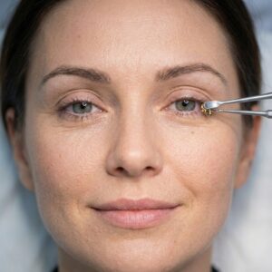
Intralesional steroid injection is a common interventional treatment for barley in the eye. This involves injecting a corticosteroid, such as triamcinolone, directly into the lesion. The steroid reduces inflammation and shrinks the barley in the eye for several days to weeks. This procedure is minimally invasive and can be performed on an outpatient basis. Patients may experience slight discomfort during the injection, and there is a small risk of side effects, including depigmentation of the skin at the injection site.
Cut and heal is another effective interventional treatment for barley in the eye. This minor surgical procedure involves making a small incision on the inside of the eyelid and draining the contents of the eyeball. The procedure is usually performed under local anesthesia and includes a short recovery period. Post-operative care includes the use of antibiotic ointment to prevent infection and warm compresses to aid healing. Complications are rare but can include bleeding, infection and scarring.
For patients with recurrent barley, treatment of underlying conditions such as meibomian gland dysfunction or blepharitis is essential. In some cases, systemic medications or long-term maintenance treatments may be required to prevent recurrence. Referral to a specialist, such as an ophthalmologist with expertise in eyelid disorders, may be necessary in complex or recurring cases.
As medical technology advances, new and innovative treatments are being tested to treat barley in the eye. These emerging and alternative treatments aim to provide effective treatment options with minimal invasiveness and improved patient outcomes.
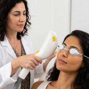
Laser treatment is one of the emerging treatments for barley in the eye. A laser can be used to make precise cuts, while reducing the risk of bleeding and infection. Laser treatment can also target and reduce the inflammation associated with styes in the eye, promoting faster resolution. Although not yet widely adopted, initial studies have shown promising results, and ongoing research continues to refine this technique.
Non-surgical techniques, such as light therapy (IPL), are also being studied for their potential in treating styes. IPL therapy involves the use of high-intensity light to heat the secretions trapped in the meibomian glands, making it easier to drain them. This treatment is already used to treat other eyelid conditions, such as meibomian gland dysfunction and rosacea, and may offer a non-invasive option for the treatment of barley in the eye.
Homeopathic remedies and herbs are alternative treatments that some patients may consider. While these treatments lack strong scientific evidence, they are often sought after by those who prefer natural approaches. Common homeopathic treatments include warm compresses with herbal infusions, such as chamomile or calendula, which have anti-inflammatory properties. Patients should be advised to use these medications with caution and under the guidance of a healthcare provider to avoid possible side effects.
While barley on the eye is usually benign, complications can arise, especially if they are not managed properly. One of the most common complications is the development of a secondary infection. When barley in the eye becomes infected, it becomes an acute hordeolum characterized by redness, sensitivity and increased swelling. Management of this complication involves the use of systemic or topical antibiotics and may require drainage if an abscess forms.
Another possible complication is the recurrence of barley in the eye. Patients with underlying conditions such as chronic blepharitis, meibomian gland dysfunction, or rosacea are at higher risk of recurrent stye. To manage this, it is essential to treat the underlying condition. Long-term strategies may include regular eyelid hygiene, use of warm compresses, and in some cases, oral medications such as doxycycline to reduce inflammation of the glands and improve meibomian gland function.
A large or untreated stye can also lead to more significant complications, such as eyelid deformity or corneal astigmatism. When barley in the eye exerts prolonged pressure on the cornea, it can cause astigmatism, resulting in blurred vision. Surgical intervention may be necessary to relieve pressure and restore normal eyelid anatomy. Post-operative care includes monitoring signs of recurrence and ensuring proper wound healing.
Patients should be educated about the signs of complications and the importance of seeking timely medical intervention. Early treatment can prevent the escalation of complications and promote a faster recovery.

Patient education and counseling are crucial elements in the management of barley in the eye to ensure effective treatment and prevention of recurrence. Educating patients about the nature of barley in the eye, its causes and treatment options helps them understand their condition and comply with the prescribed treatments.
It is essential to inform patients about the importance of maintaining good eyelid hygiene. This includes regular cleaning of the eyelid margins with a mild cleanser or eyelid scrub to prevent blockage of the meibomian glands. Demonstrating proper techniques for applying warm compresses can also be helpful. Patients should be advised to use a clean, warm washcloth and apply it to the affected eyelid for 10-15 minutes, several times a day.
Patients should be advised of the potential need for prolonged or repeated treatments, especially if they have underlying conditions such as blepharitis or meibomian gland dysfunction. An explanation of the role of these conditions in the development of barley in the eye can motivate patients to adhere to long-term management strategies.
For those undergoing interventional treatments, it is essential to provide detailed information about the procedures, potential risks and post-operative care. Patients should be aware of signs of complications, such as infection or significant changes in vision, and know when to seek immediate medical attention.
Addressing patient concerns and expectations is also important. Some patients may be concerned about the appearance of their eyelid or the potential for recurrence. Reassuring them about the effectiveness of treatment options and the availability of additional interventions if needed can help ease anxiety.
Finally, providing written instructions and resources, such as brochures or links to familiar websites, can reinforce verbal education and serve as a reference for patients after their consultation.
The treatment of barley in the eye is evolving with advances in technology and medical research, offering new and innovative approaches to management. Future directions in the treatment of barley in the eye focus on improving efficiency, reducing invasiveness and preventing recurrence.
One promising area of research is the use of laser therapy. Laser technology can provide precise and minimally invasive treatment for barley in the eye, may reduce the risk of complications and improve healing times. Studies are ongoing to determine the most effective laser parameters and protocols for the treatment of barley in the eye.
Light therapy (IPL) is another new option that shows potential. IPL therapy, already used for conditions such as meibomian gland dysfunction and rosacea, may help treat styes by reducing gland blockage and inflammation. Clinical trials are needed to establish its effectiveness and safety for barley in the eye specifically.
Gene therapy and advanced drug treatments are being researched to treat the underlying causes of stye. Research into the genetic and molecular mechanisms of meibomian gland dysfunction may lead to targeted therapies that prevent gland blockage and reduce the incidence of cataracts in the eye.
Prevention strategies are also a focus of future research. Identifying risk factors and early markers for the development of cataracts can help create preventive measures, such as personalized eyelid hygiene regimens or preventive treatments for high-risk individuals.
As the understanding of barley in the eye and its underlying causes continues to grow, healthcare providers can expect more effective and more patient-friendly treatment options. Maintaining these advances and incorporating new evidence-based methods will be essential in providing the best care for patients with ocular barley.
Barley of the eye is a common but often manageable eyelid condition that, while usually benign, can cause significant discomfort and cosmetic concerns for patients. Understanding the anatomy and pathophysiology of the meibomian glands is essential for accurate diagnosis and effective treatment. The clinical presentation usually includes localized painless swelling that can be distinguished from other eyelid lesions through careful examination.
Initial treatment with conservative treatments such as warm compresses, eyelid hygiene, and topical medications can be very effective, especially for small or less persistent styes. However, interventional treatments such as intrawound steroid injections and incision and healing provide vital options for cases unresponsive to conservative measures. These interventions can offer rapid relief and are usually well tolerated.
Emerging treatments, including laser therapy and light therapy, represent exciting advances in the field, offering less invasive and more effective alternatives. Research into genetic and molecular mechanisms, as well as innovative drug delivery systems, promises to improve future treatment options and prevention strategies.
Management of complications and recurrence requires a comprehensive approach that includes addressing underlying conditions such as blepharitis and meibomian gland dysfunction. Patient education and counseling play a central role in ensuring compliance with treatment regimens and achieving successful outcomes.
Looking forward, continued research and technological advances will likely provide even more refined and effective treatments for barley in the eye, improving patient care and reducing the burden of this condition. By staying informed and adopting new evidence-based practices, healthcare providers can ensure they are offering the best care to their patients, while fostering better health and quality of life.
סיבוכים בהרמת עפעפיים נחשבים לרוב לנדירים כאשר הניתוח מבוצע על
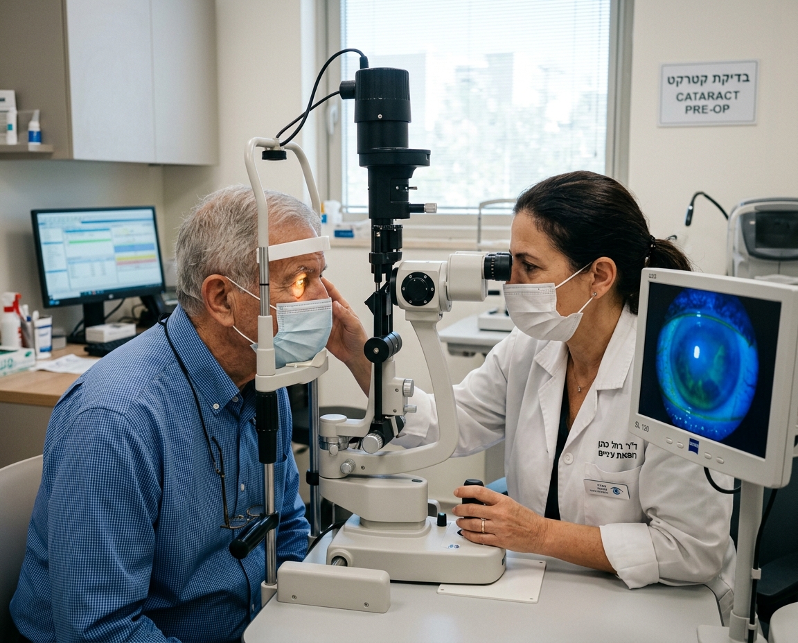
בדיקת דמעות לפני קטרקט היא שלב חיוני בהכנה לניתוח, משום
דלקת עפעפיים כרונית היא מצב דלקתי ממושך של שולי העפעפיים,